Wednesday, April 27, 2016

Sheep Eye Dissection Analysis


Displaying IMG_1949.JPG
In this lab, we got to discover the anatomy of the eye by choosing the closest thing that resembles a human eye, dissecting a sheep's eye. In just looking at the eye itself, we got to identify the the eye-lid, cornea, sclera, and the fatty tissue. The cornea controls and focuses the entry of light into the eye, and the sclera protects the eye from damage. Next, we observed the optic nerve, which transfers visual information from the retina to the vision centers of the brain.


Displaying IMG_1949.JPG


Displaying IMG_1949.JPG

After cutting the eye along the sclera, we got a view of the retina- receives light and converts the light into neural signals- which lines the posterior side of the eye. Inside of the eye contains a transparent fluid, vitreous humor, fills the cavity of the eye. This fluid, along with the aqueous humor, help in maintaining the shape of the eye.


The choroid has a large amount of blood vessels that bring nourishment and oxygen to the itself, the retina, and the sclera. Tapetum lucidum, while not being in a human eye, appears in the sheep eye, as it is used in helping animals with night vision by reflecting light at low intensities.


After removing the vitreous humor, the lens was then revealed as well as the ciliary body and the suspensory ligament. A lens is held in place by the suspensory ligament that join with the smooth muscle, that contains the ciliary body. When the smooth muscle fibers contract, it causes the lens to flatten and the degree of bending light, as a result, is reduced.


Then, we removed the lens and were able to see light coming through a oval shaped transparent opening, known as the pupil, found in the center of the iris. In contrast to a circular pupil, the sheep's pupil is oval shaped. In the second cavity between the iris and the cornea, is filled with a fluid called aqueous humor. 

Thursday, April 14, 2016

Clay Brain

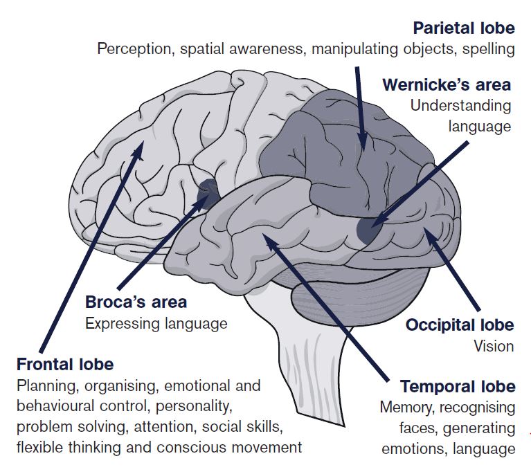
For this activity, we build a two dimensional brain out of play dough and labeled specific parts. The larger brain represents the inside view with the brain stem, cerebrum, corpus colossum, cerebellum, and the thalamus. In the smaller brain, we showed the outer portion of the brain which also represented the lobes, such as the frontal, parietal, temporal, and occipital lobe.

Wednesday, April 13, 2016

"The Woman With The Hole In Her Brain"

In the article "The Woman With The Hole In Her Brain", describes a woman who upon receiving a CAT scan discovered that a small part of her brain- the cerebellum- was missing. In the space where her cerebellum should be, was cerebrospinal fluid- which provides defense for the brain against disease. The cerebellum represents ten percent of the brains total volume; however, makes up fifty percent of its neurons. The main function of the cerebellum is controlling voluntary movements and balance, and possibly involved in the ability for a individual's motor actions and speech. Without a cerebellum, many possible complications could arise such as mental impairment, epilepsy, or a build up of fluid in the brain. The woman, however only seemed to suffer from mild speech problems and mild motor deficiency.

Frontal Lobe Damage

The frontal lobe is considered the emotional control center and the "home" of an individuals personality. It is also involved in motor function, problem solving, memory, language, impulse control, and social and sexual behavior. The frontal lobe is very vulnerable due to the fact that it is located in the front of the skull and its large size. If a individual injures his/her frontal lobe, it can effect the personality of the individual, as well as difficulty interpreting feedback from the environment, and difficulty with different facial expressions and problem solving. Even though the damage to the frontal lobe can be permanent, rehabilitation is possible to regain a small degree of prior function.